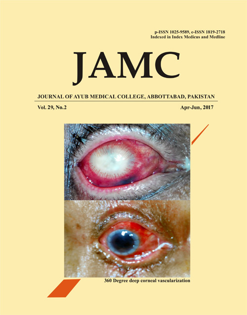
360 DEGREE DEEP CORNEAL VASCULARIZATION IN A CASE OF ENDOPHTHALMITIS WITH CORNEAL ABSCESS
Abstract
A 65-year-old male presented to us with symptoms of severe pain in right eye with loss of vision for 1 week. He had undergone a manual small incision cataract surgery elsewhere 2 weeks back. 48–72 hours post-surgery, his vision started decreasing and he presented to us in a state shown in the figure 1. On examination, a provisional diagnosis of post-operative endophthalmitis was made. The anterior segment showed ciliary congestion, 360º corneal vascularization with corneal abscess. Corneal neovascularization (or simply corneal vascularization) results from the growth of new vessels from the limbal vasculature into the normally avascular cornea.1 Corneal vascularization may be superficial or deep. In superficial variety, the vessels are arranged in an arborizing pattern beneath the corneal epithelium and their continuity with conjunctival vessels can be traced.2 In deep vascularization, vessels are usually straight, do not anastomose and their continuity beyond limbus cannot be traced.2 Deep vessels can be arranged as terminal loops(type A), brush(type B), parasol, umbel, network or as interstitial arcade.2 Common causes of deep vascularization are interstitial keratitis, disciform keratitis, deep corneal ulcer, chemical burns, etc.2 Treatment options include topical corticosteroids, radiation, diathermy and peritomy but none is satisfactory for the deep variety. Our patient underwent a 360º peritomy followed by therapeutic penetrating keratoplasty (TPK) and vitrectomy. The status post-management is shown in figure-2. This pictorial aims to demonstrate two types of deep corneal vascularization seen in same patient, which is quite rare. Corneal vascularization can lead to loss of corneal transparency and hence needs management energetically.References
Giulio F, Chiara G, Paolo R (2015) Corneal Neovascularization: A Translational Perspective. J Clin Exp Ophthalmol 6:387. doi:10.4172/2155-9570.1000387
Khurana AK. Comprehensive ophthalmology. 4th ed. New Delhi: New Age International; 2007.
Published
Issue
Section
License
Journal of Ayub Medical College, Abbottabad is an OPEN ACCESS JOURNAL which means that all content is FREELY available without charge to all users whether registered with the journal or not. The work published by J Ayub Med Coll Abbottabad is licensed and distributed under the creative commons License CC BY ND Attribution-NoDerivs. Material printed in this journal is OPEN to access, and are FREE for use in academic and research work with proper citation. J Ayub Med Coll Abbottabad accepts only original material for publication with the understanding that except for abstracts, no part of the data has been published or will be submitted for publication elsewhere before appearing in J Ayub Med Coll Abbottabad. The Editorial Board of J Ayub Med Coll Abbottabad makes every effort to ensure the accuracy and authenticity of material printed in J Ayub Med Coll Abbottabad. However, conclusions and statements expressed are views of the authors and do not reflect the opinion/policy of J Ayub Med Coll Abbottabad or the Editorial Board.
USERS are allowed to read, download, copy, distribute, print, search, or link to the full texts of the articles, or use them for any other lawful purpose, without asking prior permission from the publisher or the author. This is in accordance with the BOAI definition of open access.
AUTHORS retain the rights of free downloading/unlimited e-print of full text and sharing/disseminating the article without any restriction, by any means including twitter, scholarly collaboration networks such as ResearchGate, Academia.eu, and social media sites such as Twitter, LinkedIn, Google Scholar and any other professional or academic networking site.


