
EPIPLOIC APPENDAGITIS
Abstract
A 57-year-old female presented to the emergency department with a two-day history of left lower quadrant abdominal pain. Vital signs and results of a complete blood count and basic metabolic panel were unremarkable. Abdominal exam was significant for localized left lower quadrant peritonitis. Computed tomography of the abdomen and pelvis showed diffuse fatty infiltration and mild peritoneal thickening in the left abdomen suggestive of omental infarct/fat necrosis (Figure-1, arrow). Patient was treated conservatively using NSAIDS with no success. Laparoscopy revealed large infarcted epiploic appendage adhered to the anterior abdominal wall along with the greater omentum (Figure-2, arrow). After the epiploic appendage was separated from the anterior abdominal wall and greater omentum, torsion of its vascular pedicle was seen (Figure-3, arrow). The vasculature was divided with laparoscopic electrocautery device and the specimen was removed from the abdomen (Figure-4). Her pain improved immediately following the surgery. She was discharged home the next day.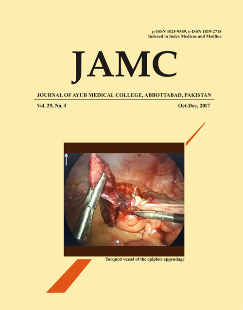
Published
Issue
Section
License
Journal of Ayub Medical College, Abbottabad is an OPEN ACCESS JOURNAL which means that all content is FREELY available without charge to all users whether registered with the journal or not. The work published by J Ayub Med Coll Abbottabad is licensed and distributed under the creative commons License CC BY ND Attribution-NoDerivs. Material printed in this journal is OPEN to access, and are FREE for use in academic and research work with proper citation. J Ayub Med Coll Abbottabad accepts only original material for publication with the understanding that except for abstracts, no part of the data has been published or will be submitted for publication elsewhere before appearing in J Ayub Med Coll Abbottabad. The Editorial Board of J Ayub Med Coll Abbottabad makes every effort to ensure the accuracy and authenticity of material printed in J Ayub Med Coll Abbottabad. However, conclusions and statements expressed are views of the authors and do not reflect the opinion/policy of J Ayub Med Coll Abbottabad or the Editorial Board.
USERS are allowed to read, download, copy, distribute, print, search, or link to the full texts of the articles, or use them for any other lawful purpose, without asking prior permission from the publisher or the author. This is in accordance with the BOAI definition of open access.
AUTHORS retain the rights of free downloading/unlimited e-print of full text and sharing/disseminating the article without any restriction, by any means including twitter, scholarly collaboration networks such as ResearchGate, Academia.eu, and social media sites such as Twitter, LinkedIn, Google Scholar and any other professional or academic networking site.


