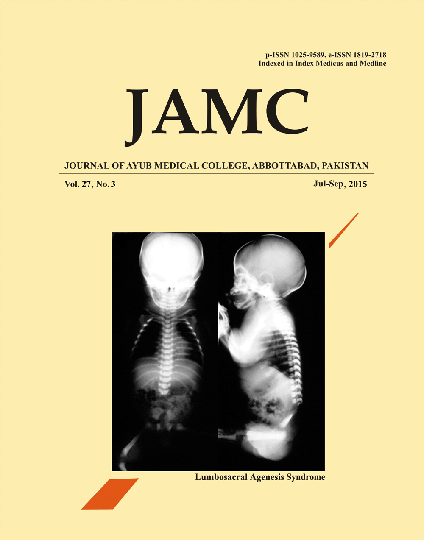
LUMBOSACRAL AGENESIS SYNDROME IN A BABY BORN TO DIABETIC MOTHER
Abstract
This newborn was delivered full term normal vaginal delivery to a mother who had uncontrolled Diabetes. Baby had bilateral hypotonia in lower limbs and a sacral dimple. X-ray show absent vertebrae below 2nd lumber. Lumbosacrococcygeal agenesis is one of the complications of uncontrolled Maternal Diabetes. In general population the incidence of this condition is less than 0.01%.1 16% of the patients are infants of Diabetic mothers. Infants of less than 1% of diabetic mothers have this or any other osseous deformity. LSCA is graded in four types according to the severity of the condition.Type1 where only one side of the sacral agenesis is found producing hemevertebrae. Type II with complete bilateral agenesis of sacrum. Type III is total Saccrococcygeal agenesis with variable Lumber agenesis and type IV with total saccrococcygeal agenesis with variable lumber agenesis and last formed vertebrae resting on ilial bone.2,3 According to this classification our patient was having Type-III deformity. Other problems in IDM babies are hypoglycemia, Hypocalcemia, Polycythemia, cardiac malformations ,short colon syndrome. Management depends on the abnormal findings in the lower limbs and urinary symptoms. Prophylactic Antibiotics to prevent urinary Tract infections, catheterization to prevent distended bladder and laxatives for constipation may be needed. Physiotherapy for the lower limbs is important.References
Diel J, Ortiz O, Lasada RA, Price DB, Hayt MW, Katz DS. The sacrum: pathologic spectrum, multimodality imaging and subspecialty approach. RadioGraphics 2001;21:83–104.
Gudinchet F, Maeder P, Laurent T, Meyrat B, Schnyder P. Magnetic resonance detection of myelodysplasia in children with Currarino triad. Pediatr Radiol 1997;27:903–4.
Rensahw TS. Sacral agenesis: a classification and review of 23 cases. J Bone Joint Surg 1978;60(A):373–83
Published
Issue
Section
License
Journal of Ayub Medical College, Abbottabad is an OPEN ACCESS JOURNAL which means that all content is FREELY available without charge to all users whether registered with the journal or not. The work published by J Ayub Med Coll Abbottabad is licensed and distributed under the creative commons License CC BY ND Attribution-NoDerivs. Material printed in this journal is OPEN to access, and are FREE for use in academic and research work with proper citation. J Ayub Med Coll Abbottabad accepts only original material for publication with the understanding that except for abstracts, no part of the data has been published or will be submitted for publication elsewhere before appearing in J Ayub Med Coll Abbottabad. The Editorial Board of J Ayub Med Coll Abbottabad makes every effort to ensure the accuracy and authenticity of material printed in J Ayub Med Coll Abbottabad. However, conclusions and statements expressed are views of the authors and do not reflect the opinion/policy of J Ayub Med Coll Abbottabad or the Editorial Board.
USERS are allowed to read, download, copy, distribute, print, search, or link to the full texts of the articles, or use them for any other lawful purpose, without asking prior permission from the publisher or the author. This is in accordance with the BOAI definition of open access.
AUTHORS retain the rights of free downloading/unlimited e-print of full text and sharing/disseminating the article without any restriction, by any means including twitter, scholarly collaboration networks such as ResearchGate, Academia.eu, and social media sites such as Twitter, LinkedIn, Google Scholar and any other professional or academic networking site.


